Infections caused by anaerobic bacteria are common, and may be serious and life-threatening. Anaerobes predominant in the bacterial flora of normal human skin and mucous membranes, and are a common cause of bacterial infections of endogenous origin. Infections due to anaerobes can evolve all body systems and sites. The predominate ones include: abdominal, pelvic, respiratory, and skin and soft tissues infections. Because of their fastidious nature, they are difficult to isolate and are often overlooked. Failure to direct therapy against these organisms often leads to clinical failures. Their isolation requires appropriate methods of collection, transportation and cultivation of specimens. Treatment of anaerobic bacterial infection is complicated by the slow growth of these organisms, which makes diagnosis in the laboratory only possible after several days, by their often polymicrobial nature and by the growing resistance of anaerobic bacteria to antimicrobial agents.

The site is made of a home page that presents new developments and pages dedicated to infectious site entities.


Clostridial Infections: Colitis, Tetanus, Botulism


Clostridial diarrhea and pseudomembranous colitis


Pseudomembranous colitis (PMC) is commonly associated with hospitalization and prior antibiotic exposure and can affect adults and children. Other risk factors include use of proton pump inhibitors (eg, omeprazole); use of nasogastric, gastrostomy or jejunostomy tubes; and underlying 

bowel disease or surgery, among others.

PMC is caused almost exclusively by toxins produced by Clostridium difficile. The clinical spectrum of this disease may range from a mild, nonspecific diarrhea to severe colitis with toxic megacolon, perforation, and death.1


Microbiology & pathogenesis

Ampicillin, amoxicillin, the second and third generation cephalosporins and clindamycin are most frequently associated with development of PMC, although nearly all antimicrobials have been implicated as causes of diarrhea and colitis.1 Antibiotics that infrequently cause PMC are penicillins, first‑generation cephalosporins, chloramphenicol, macrolides, quinolones, tetracyclines, aminoglycosides, rifampin, and trimethoprim‑sulfamethoxazole. The initial research  associated PMC with clindamycin resistant C. difficile. However, this condition was related to all other antimicrobials. 2,3 

Gram stain of Clostridium difficile 


Numerous studies2,3 implicated C. difficile as an etiologic agent of PMC . Other Clostridium (Clostridium innocuum, Clostridium oroticum, and Clostridium ramosum) were also recovered with Candida spp. and aerobic Gram-negative bacilli. Severe PMC can be associated with C. difficile toxin in the stool without previous antibiotic exposure. 8,9

C. difficile produces at least two toxins, both necessary to produce PMC, an enterotoxin (toxin A), and a cytotoxin (toxin B). The cytotoxin is potent in tissue culture assays and is a sensitive and specific marker for C. difficile-induced disease, whereas toxin A is more potent in biologic assays in animal models, and may be more important in gastrointestinal complications.

Most stools from patients with PMC contain the C. difficile organism and its` cytotoxin. Individuals treated with antibiotics can acquire the organism from an environmental source. A third of healthy newborns harbor C. difficile in their stool with detectable toxin without clinically apparent consequences.4  Carrier rates for C. difficile in stool decrease with age. C. difficile can be spread in a neonatal nursery, hospital wards and households, by hands of personnel, and by fomites. 3
C. difficile ability to produce PMC mostly in the presence of antibiotic exposure is explained by its` enhanced growth in an environment in which there is reduced bacterial competition.

C. perfringens, a producer of a exotoxin can cause toxigenic diarrhea. C. perfringens diarrhea has been associated with ingestion of contaminated beef, beef products, and poultry. C. perfringens produces a thermolabile toxin, which is synthesized before ingestion prior to sporulation and in the vegetative phase in the gastrointestinal tract. It effects the proximal small bowel by activating intestinal adenyl cyclase, resulting in increased intestinal fluid secretion and decreased reabsorption.

Specimen of psudomembranous colitis


Clinical presentation

Disease due to C. difficile represents a wide spectrum- asymptomatic cases, watery diarrhea, dysentery, PMC, complicated colitis and bacterial metastatic infections.5 Watery diarrhea, infrequently with blood or mucus, develops in most patients. Abdominal pain, cramps, lower quadrant tenderness, fever and leukocytosis are common. Systemic symptoms varies from mild to severe and the disease may be fatal. The duration of symptoms in mild disease ranges from 7 to 10 days after discontinuation of the antibiotic.

C. perfringens diarrhea is characterized by sudden onset,  colicky abdominal pain,  and foul stools free of blood and mucus.

Specimen of psudomembranous colitis 


Diagnosis

The diagnostic test of choice to detect toxin B is a tissue culture assay which demonstrates a cytopathic toxin that is neutralized by clostridial antitoxin.3 Several new enzyme immunoassays can detect toxin A, B, or both. Latex particle agglutination assay is not as reliable. The immunoblot assay and polymerase chain reaction, can detect toxin A only.

Stool cultures for C. difficile using selective medium should be attempted, accompanied by a reliable toxin assay. Determining the role of C. perfringes in diarrhea is difficult.  Fluorescent stained antibodies to C. perfringes capsule can be used to identify the organism in stool.

Endoscopy can detect the typical plaque like lesions of the pseudomembrane. Sigmoidoscopy may be sufficient when the distal colon is involved, but if the pseudomembranes is restricted to the right colon, colonoscopy is necessary. Findings range from a normal mucosa to erythema, edema, friability, ulceration, hemorrhage, and PMC. Air-contrast barium enema is often nonspecific.
Histologically, earliest findings are patchy necrosis of colonic epithelium with intraluminal exudation of neutrophils and fibrin. Epithelial ulcers become apparent and pseudomebranes, comprised of leukocytes, fibrin, mucus and cell debris, then cover the diffuse epithelial necrosis.


Management

Supportive therapy and discontinuance of the antibiotic therapy are generally adequate for mild diarrhea. Comparative efficacy data on metronidazole and vancomycin in adults (there are no pediatric data) from controlled trials show that these two agents are equally effective. Some data, , suggest that vancomycin may be more effective for more severe disease.Those with severe symptoms or persistent diarrhea require aggressive therapy. Vancomycin is effective, but is expense, tastes bad, and is associated with up to 20% relapse.

Cholestyramine, an anion exchange resin that binds C. difficile toxins, is an alternative to vancomycin. It is more likely to result in treatment failure but is less likely to be followed by relapse. Metronidazole and bacitracin can also be used. Metronidazole orally has similar efficacy to vancomycin orally in mild and moderate cases,  lower cost and does not select enterococcal resistance to vancomycin. Disadvantages of metronidazole are occasional resistance of C. difficile, rare induction of PMC, and its complete absorption so that bactericidal levels are erratic. Also continued use may result in neurotoxicity. Anti-motility agents, including loperamide, diphenoxylate hydrochloride with atropine, and opioids, should be avoided as they can adversely affect the ability to clear the toxins.

Fever, systemic manifestations, and severe diarrhea generally improve within one  to 2 days of therapy, but diarrhea may last for 4 to 5 days.  Relapses may be caused by re-acquisition or persistence of spores. Most patients with a relapse respond to the re-treatment, but may experience multiple recurrences. 

Options for management of multiple relapses include: vancomycin given as tapered or pulsed regimens, fidaxomicin, vancomycin or metronidazole plus Saccharomyces boulardii or Fleischmann’s baker’s yeast; or followed by cholestyramine with or without lactobacilli; intravenous immunoglobulin, solution of fresh stool from healthy donor (fecal transplant); and broth culture bacteria.
Treatment of C. perfringens diarrhea is supportive. The disorder is self-limiting and lasts less than 24 hours.
Surgical intervention may be required in severe cases unresponsive to medical therapy or to manage complications such as toxic megacolon, or colonic perforation. In fulminate PMC vigilance is necessary to detect peritonitis and abdominal cellulitis that indicate intestinal perforation.


Complications

Complications include dehydration, electrolyte imbalance, hypotension, hypoalbuminemia with anasarca, transverse volvulus, colon perforation and toxic megacolon. Severe cases are prone to secondary systemic infection because of acquired malnutrition, hypogammaglobulinemia, lymphopenia, ascites, pleural effusions and bacteremia from the intestinal inflammatory process.
Recurrent colitis and diarrhea occur in one of five patients 2 to 8 weeks after completion of therapy; occasionally, more than 6 episodes may occur.

Toxic megacolon


Prevention

Administration of antibiotics associated with PMC to patients with intestinal disease should be avoided. Antibiotic therapy should always be done prudently, and  limited to those who unequivocally need it.
Preventation of recurrences include avoidance of any antimicrobials in the first 2 months after PMC. Innovative strategies with adjunctive agents and probiotics are used in patients with recurrences.
The spread of C. difficile within the hospital should be prevented by strict adherence to hand‑washing practices and limitations on use of broad‑spectrum antimicrobial agents and unnecessary antimicrobial therapy. Strict contact isolation, cohorting patients and personnel in epidemics and, rarely, closing units may be required.

Sigmoidoscopes and colonoscopes should be decontaminated to avoid possible transmission of C. difficile. The only sporicidal agents effective against C. difficile are sodium hypochlorite  and glutaraldehydes. 


REFERENCES           

 1.  Cleary RK. Clostridium difficile-associated diarrhea and colitis: clinic manifestations, diagnosis, and treatment. Dis Colon Rectum  ;41:1435-49, 1998.
2.        Brar HS, Surawicz CM Pseudomembranous colitis: an update. Can J         Gastroenterol;14:51-6, 2000.
3.        Johnson, S., Gerding, D.N.: Clostridium difficile-associated diarrhea. Clin Infect Dis 26:1027–34; quiz 1035-6, 1998.
4.        Sherertz, R.J.,  Sarubbi, F.A.: The prevalence of Clostridium difficile and toxin in a nursery population: A comparison between patients with necrotizing enterocolitis and an asymptomatic group. J. Pediatr. 100:435-9, 1982.
5.        Feldman, R.J., Kallick M., Weinstein, M.P.,: Bacteremia due to Clostridium difficile: case report and review of extraintestinal  Clostridium difficile  infections Clin Infect Dis 20:1560-2, 1995.


Tetanus 


Tetanus is an acute toxemic illness caused by Clostridium tetani infections at a laceration or skin break, burns, puerperal infections, umbilical stump infections (tetanus neonatorum), and surgical sites (due to contaminated sutures, dressings, or plaster). 1

Epidemiology

Tetanus is a major cause of death in areas of the world without appropriate hygiene and immunization programs. The incidence of tetanus varies widely throughout the world; in the US 50 to 100 cases are reported annually. 2 Beyond the neonatal period, the attack rate and age related mortality rate is higher in males. Infected wounds, abscesses, parenteral drug abuse, major trauma, and animal-related injuries account for 25% of the tetanus-associated injuries.


Etiology and Pathophysiology

C. tetani spores resist heat and the usual antiseptics and can persist in tissues for months. They can survive in soil for years and can be found in house dust, soil, salt, fresh water, contaminated heroin and the feces of many animal species.

The portal of entry is usually the site of puncture wounds or scratches. Deep puncture wounds, burns, crushing, and other injuries that promote favorable growth conditions for anaerobic organisms may lead to the development of tetanus.

Following introduction into tissues, spores convert to vegetative forms, multiply, and elaborate tetanospasmin.  The toxin enters the peripheral nerve and travels through the nerves or is transferred by the lymphocytes to the central nervous system (CNS ).3 It effects the nervous system centrally and peripherally. It binds to gangliosides at the presynaptic nerve ending in the neuronal membrane, prevents release of neurotransmitters, and affects polarization of postsynaptic membranes in complex polysynaptic reflexes. The resulting lack of inhibitory impulses produces the characteristic spasms, seizures, and sympathetic overactivity. The toxin has no effect on the mental status, and consciousness is not impaired.

Tetanospasmin also becomes bound to gangliosides within the CNS where it suppresses the motor neurons and interneurons.Antitoxin may prevent binding in the CNS only if binding has taken place in the periphery. The length and course of the illness are determined by the location and amount of the bound toxin. Tetanus complete course ranges from 2 to 6 weeks. 
Gram stain of Clostridium tetani

Clinical Presentation

Tetanus can present as: localized, generalized, cephalic, or neonatal. 1
Generalized Tetanus is the most common form. The initial findings are trismus (“risus sardonicus”) due to spasms of the parapharyngeal and masseter muscles, pain, swallowing difficulty, and unilateral or bilateral stiffness of the neck and other muscle groups, such as those of the abdomen or thorax, resulting in severe opisthotonos. The tetanic contractions progress,  with significant worsening of symptoms. Painful spasms of contraction further contort and distort the  posture. All voluntary muscles can be effected and the disease may involve the larynx, which can be fatal. Fractures of vertebrae or other bones and hemorrhage into muscles and cardiovascular instability such as labile hypertension, and episodes of tachycardia or other tachyarrhythmias can occur. The sympathetic overactivity or superinfections, (e.g. pneumonia ) can cause fever.   Minor stimuli including light, drafts, noises or voices, and light touch may trigger spasms. Patients remain fully conscious and anxiety and pain further complicate management.

Opisthotonus


Localized Tetanus: This rare presentation occurs when circulating antitoxin prevents general toxin spread but is insufficient to stop local uptake at a wound site.  This results in weeks long steady and painful muscle contraction in the wound region.

Cephalic Tetanus: This rare manifestation exclusively involves the cranial nerves after entry of C. tetani  into wounds or chronic head and neck infections. Cranial nerve VII is most frequently involved. Cephalic tetanus may precede generalized disease.

Cephalic tetanus


Neonatal Tetanus: This generalized form develops in infants delivered vaginally to unimmunized mothers. Birth practices, such as applying mud or feces to the umbilical stump, increase the risk of acquiring this illness. The mortality rate is high, with infants dying of CNS hemorrhage, pneumonia, pulmonary hemorrhage, and laryngeal spasms.


Child with tetanus


Neonatal tetanus


Diagnosis and Differential Diagnosis

Tetanus is rare in developed nations. Conditions that can mimic tetanus include parapharyngeal and peritonsillar abscesses, poliomyelitis and other forms of viral encephalomyelitis, meningoencephalitis (including rabies), Bell's palsy, hypocalcemic tetany, and dystonic reactions to phenothiazines. 4

Specific diagnosis of tetanus by routine laboratory tests is difficult. Gram stains and anaerobic cultures of wounds can reveal the characteristic Gram-positive bacilli with terminal spores.


Management and Prognosis

Appropriate treatment based on clinical diagnosis is warranted even without specific confirmatory laboratory tests. The goals of therapy are to eradicate C. tetani, neutralize its toxin, and  provide supportive care. Specific therapy includes IM administration of tetanus immune globulin (TIG) to neutralize circulating toxin before it binds to neuronal cell membranes. Early administration may prevent toxin spread to the CNS. The recommended dosage of TIG ranges from 500 to 3000 U. The newborn dose is TIG (250 U).

Additionally specific therapy includes antimicrobial agents for C. tetani such as penicillin G administered as 50,000 U/kg IV q6h for 10 days. Alternatives for those allergic to penicillin include tetracycline (40 mg/kg/day; maximum of 2 g) or IV vancomycin (30 to 40 mg/kg/day).
Wound are left open and are cared by debridement, irrigation, and foreign bodies removal.
Patients should be managed in an intensive care setting in a quiet darkened room, utilizing suction , oxygen, cardiac and respiratory monitors, a ventilator, and tracheostomy equipment. Sedation and muscle relaxation should be instituted, usually with diazepam ( 0.1 to 0.2 mg/kg IV ,q 4 to 6 h). Additional sedation with phenothiazines and therapeutic paralysis may be needed. Patients who undergo therapeutic paralysis must be sedated to avoid anxiety.
Neuromuscular blockade can be achieved with curariform drugs: Vecuronium ( an initial dose of 0.08 to 0.1 mg/kg IV, with maintenance doses of 0.01 to 0.15 mg/kg q 30 to 60 m), and Doxacurium, ( initial dose is 0.03 to 0.05 mg/kg IV, followed by 0.01 mg/kg in 60 to 90 m). Hypertension may require treatment with beta-blocking agents. ( i.e. propranolol 0.01 to 0.10 mg/kg q 6 to 8 hr, for 2 - 3 w).

Maintenance of adequate nutrition, hydration,skin care, and excretory functions is of outmost importance. Patients must be immunized with tetanus toxoid to prevent further disease.

Complications
These include those due to direct toxicity (laryngeal and phrenic nerves palsy, and cardiomyopathy), secondary to spasms  (respiratory compromise, rhabdomyolysis, myositis ossificans circumscripta, and vertibral compressed fracture), respiratory compromise (hypoxic cerebral injury), rhabdomyolysis (acute renal failure), and psychological.

Prevention

Active immunization with tetanus toxoid is the most effective mean of protection. The primary series of tetanus toxoid, administered as DTP vaccine at 2, 4, and 6 m and a booster 12 months later, ensures protection. Additional boosters are given each decade, with further tetanus prophylaxis after acute wounds, as advocated by the American Academy of Pediatrics Advisory Committee on Immunization Practices (Table 1).5

Individuals who have full primary immunization and appropriate boosters need no tetanus prophylaxis beyond local wound care for clean minor wounds but should receive a toxoid booster after a tetanus-prone injury if the most recent dose was received more than 5 y previously. Patients who are not known to have completed the primary series require a tetanus toxoid booster after any penetrating wound and TIG after a tetanus-prone injury. The prophylactic dose of IM TIG is 250 to 500 U.


TABLE 1 . Tetanus prophylaxis in wound management

Type of wound
Immunization history

Clean, mirror

All others

Three or more doses of tetanus toxoid
No TIG; toxoid only if > 10 yr since last dose
No TIG; toxoid only if > 5 yr since last dose
Fewer than three doses or uncertain history
No TIG;
 toxoid, 0.5 mL
TIG; 500 units; toxoid, 0.5 mL
* Equine tetanus antitoxin should be used when TIG is not available.
TIG = tetanus immune globulin.


References

1.        Brook I. Current concepts in the management of Clostridium tetani infection.                      Expert Rev Anti Infect Ther. 2008 :327-36.
2.         Bardenheier, B., Prevots, D.R., Khetsuriani, N., et al.: Tetanus surveillance-united states, 1995-1997. Mor Mortal Wkly Rep CDC Surveill Summ 47:1-13, 1998.
3.         Montecucco, C., Schiuvo, G.: Mechanism of action of tetanus and botulinium neurotoxin. Mol. Microbiol 13:1-8. 1994.
4.         Henderson, S., Mody, T., Groth, D.E. et al.: The presentation of tetanus in an emergency department. J. Em. Med 16:705-8. 1998.
5.         American Academy of Pediatrics. Tetanus (lockjaw).Pickering L K editor. 2009;665-660. Red Book: Report of the Committee on Infectious Diseases, 28th ed. Elk Grove Village, IL: American Academy of Pediatrics; 2009.


BOTULISM


Botulism is caused by a neurotoxin produced from the anaerobic, spore-forming bacterium Clostridium botulinum. Botulism in humans is usually caused by toxin types A, B, and E.
These are forms of botulisms: food-borne botulism, wound botulism, infant botulism, adults intestinal toxemia botulism, inhalation botulism,  unclassified botulism (in patients over 12 month old with proven botulism without an ingestion source), and iatrogenic botulism. Symptoms and pathologic findings relate to the toxin’s effects on the nervous system and are characterized by neuromuscular dysfunction and resultant flaccid paralysis of muscles.

FOOD-BORNE BOTULISM

Epidemiology

Food-borne botulism, the most common form of botulism, usually occurs in small sporadic outbreaks.1 Botulism toxin is the most dangerous toxin known, as only 0.5 g of toxin A is lethal for humans. The toxin is ingested with food that has become contaminated with C. botulinum during canning or other preparatory process.1

C. botulinum spores are highly heat-resistant; they may survive several hours at 100°C; however, exposure to moist heat at 120°C for 30 minutes will kill the spores. The toxins, on the other hand, are destroyed by heat, and cooking food at 80°C for 30 minutes safeguards against botulism.1 As a result, the ingestion of preformed toxin, not simply spores, is required in adult botulism. In infant botulism, however, the toxin can be produced by incubation of the spores within the gut.2
Most U.S. outbreaks of botulism are associated with food products that are not heated adequately before consumption and in which spores form toxins. In the United States, preserved foods in which the toxin is most commonly found include string beans, corn, mushrooms, spinach, olives, onions, beets, asparagus, seafood, pork products, and beef.2,3 Improperly smoked or canned fish is the source of type E intoxications. Botulinum spores are common in soil, dust, lakes, and other environmental matter and can contaminate fruits, vegetables, meats, and fish. Honey has become recognized as a potential source of C. botulinum spores and one of the botulism.

Canned food contaminated with Clostridium botulinum


Etiology and pathophysiology

The causative agent is one of several eight closely related serologically toxins elaborated by the sporulating, C. botulinum. Human poisoning usually is caused by type A, B, or E toxin, rarely by type C1, C2, D, F or G.4

Three steps are necessary for toxin-induced neuromuscular blockade: (a) transport across the intestinal wall into the serum, (b) binding to neuronal receptors, and (c) internalization of bound toxin, an irreversible step leading to impairment of neurotransmitter release and resultant neuromuscular blockade.4

Botulinal toxin type A has therapeutic value in the therapy of a several neurologic and ophthalmologic disorders through chemical denervation.5 They are used as a therapeutic agent through local instillations in strabismus, blepharospasm, and other facial nerve disorders.

Clostridium botulinum


Clinical findings

There are four cardinal clinical features of botulism6:
1.         Symmetric and descending neurologic manifestations.
2.         Intact mental processes.
3.         No sensory disturbances, although vision may be impaired because of extraocular muscle involvement.
4.         Absence of fever unless secondary infectious complications occur.

At 18 to 48 hours after ingestion of tainted food, patients present with cranial nerve dysfunction manifested by diplopia, dysphagia, and difficulty in speaking. They remain lucid, although anxiety and agitation may develop. Additional signs may include pupillary dilation, vertigo, tinnitus, and dry mouth and mucous membranes. The descending progression of paralysis in botulism occurs at various rates, spreading and involving muscles of respiration and most voluntary musculature. The major manifestation is respiratory embarrassment, which may appear gradually or suddenly. 2,6 

Involvement of the gastrointestinal tract varies and is related somewhat to the toxin serotype. Types A and B, cause abdominal complaints (e.g., abdominal pain, bloating, cramps, diarrhea) in approximately one-third of patients that are replaced quickly by constipation. Type E produces more significant gastrointestinal complaints than do the other types. 2,6

The chief cause of mortality is respiratory or bulbar paralysis. The typical duration of symptoms exceeds 1 month, and full recovery from weakness and fatigability may require as long as 1 year.


Diagnosis

Diagnosis is suggested by the pattern of neuromuscular disturbances and a likely food source and is confirmed by demonstration of botulinus toxin or C. botulinum in suspected food, vomitus, and serum. Electromyography (EMG) can be helpful in diagnosis when lowered amplitude action potentials following low-frequency stimulation and posttetanic facilitation of the muscle action potential can be demonstrated.

The isolated muscle action potential is reduced, but repetitive nerve stimulation results in facilitation of the action potentials.

Suspected cases must be reported to local and state health authorities and to the Center for Disease Control and Prevention  (tel. 800-232-4636), from which trivalent antitoxin for types A, B, and E is available. Blood and stool samples should be transported to a laboratory (usually state health departments) equipped to determine botulinal toxin. Stool should be cultured for Clostridium because C. botulinum . Definitive diagnosis can be made by demonstration of preformed toxin by the mouse inoculation test.

Botulism may be confused with poliomyelitis, viral encephalitis, myasthenia gravis, Guillain-Barré syndrome, tick paralysis, and atropine or mushroom poisoning.


Management

Mortality rate has dropped from 60% to 16%, most likely a result of improvement in critical care management. The longer incubation period, the better is the prognosis.
Management involves optimal supportive care and specific therapy directed at neutralizing unbound toxin, eradicating any infection with C. botulinum and management of secondary infections. Speed is essential in diagnosis so that circulating toxin can be neutralized before it binds to nerve endings. Induced vomiting and gastric lavage is carried out if exposure has occurred within several hours. Bowel purges have been suggested. Airway control, proper oxygenation and management of adequate ventilation is of great importance. Endotracheal intubation may be required. 7

Oral or parenteral antimicrobials such as penicillin have limited value but may destroy some viable C. botulinum organisms. Aminoglycosides can affect the neuromuscular junction and potentiate the effect of botulinal toxin and should be avoided.

The administration of appropriate antitoxin without delay is recommended. It has little effect with types A and B and a greater effect with type E toxin poisoning. Several forms of equine botulism antitoxin are available: monovalent type E, bivalent AB, trivalent ABE, and polyvalent ABCDEF. If the causative toxigenic type is unknown, or if type-specific antitoxin is not available, the trivalent ABE preparation should be used. 7 

If hypersensitivity to horse serum is not demonstrated, the antitoxin can be given intravenously at a dose of one vial every four hours for a total of four or five vials. If toxin is still detectable in subsequent serum samples, additional antitoxin may be given after repeat skin testing. Guanidine may enhance the release of acetylcholine from nerve terminals and may help in mild cases; however, guanidine is less effective in overcoming respiratory muscle paralysis.


Prevention

Prevention is of utmost importance. Proper home and commercial canning and adequate heating of food before serving are essential. Food showing any evidence of spoilage should be discarded. After a case has been recognized, health authorities must be notified so that other potential cases can be identified and treated expectantly.


WOUND BOTULISM

Epidemiology

Wound botulism is rare in the USA. Most cases involve wounds located on an extremity.


Predisposing factors

Wound botulism is associated with major soil contamination through compound fractures, severe trauma, lacerations, puncture wound, “skin popping” of black tar heroin, and hematoma.7


Etiology

The pathogenesis and etiology of wound botulism are similar to the food-borne disease. Most wound cases have been associated with type A toxin-producing organism, and some with type B.


Clinical signs and diagnosis

The incubation period is longer (4 to 18 days) than for food-borne illness (six hours to eight days). The clinical manifestations are similar to those of food-borne botulism except for the lack of early gastrointestinal symptoms. The wound can look benign, with minimal erythema, induration or discharge, but the organism and toxin are usually present.2
The lesion is unroofed to obtain specimens for culture and toxin assay. Diagnosis is confirmed by EMG and demonstration of toxin in serum or by isolation of C. botulinum and/or toxin from the wound in association with appropriate clinical findings.

Wound botulism 


Management

Treatment of wound botulism must include debridement, drainage, and wound irrigation. Good supportive care, as previously described, primarily respiratory support, is also an important aspect of management. Although antitoxin will not improve paralysis from toxin already bound at the neuromuscular junction, antitoxin will bind circulating toxin.


Adult intestinal toxemia botulism
    
Adult intestinal toxemia botulism occurs rarely and sporadically and results from of absorption of toxin produced in situ by  botulinum-toxin producing Clostridia that colonizes the intestine. Generally, patients have an anatomical or functional bowel abnormality or are using antimicrobials, which may select fastidious Clostridia species from the normal bowel flora. 8-11 The symptoms may be protracted and relapse even after  treatment with antitoxin because of the ongoing intraluminal production of toxin. Typically there is no known food or wound source and prolonged excretion of organisms and toxin are present in the stool.

Inhalational botulism.    
Inhalational botulism is iatrogenic and was described only once among laboratory workers in 1962, with symptoms similar to foodborne botulism. 12 Deliberate dissemination of botulinum toxin by aerosol  is a potential biological weapon could produce an outbreak of inhalational botulism. 13
     
Iatrogenic botulism 
  
Iatrogenic botulism occurs after injection of botulinum toxin for therapeutic or cosmetic purposes. The doses recommended for cosmetic treatment are too low to cause systemic disease. However, higher doses injected for treatment of muscle movement disorders have caused systemic botulism-like symptoms in patients. 13 Injection of highly concentrated botulinum toxin have caused severe botulism in some patients who received it for cosmetic purposes.
 Iatrogenic botulism was reported in four patients who received botulinum toxin injections using an unlicensed, highly concentrated preparation of botulinum toxin A. 14   The patients received doses as high as 2857 times the human lethal dose by injection and had serum toxin levels 21 to 43 times the estimated lethal human dose by injection. All patients survived following administration of equine serum antitoxin, and required prolonged mechanical ventilation and physical rehabilitation.


INFANT BOTULISM


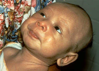
Infant botulism occurs as a result of absorption of the heat labile neuro-toxin that is produced by that colonizes the intestinal tract of an infant younger than one year. It is an age limited neuro-muscular illness that is different from the classical illness in that the toxin is generated by the organisms in the infant's intestine, the thereafter absorbed to produce the illness.


The organisms generally are squired from the environment and include honey and soil. Clinical manifestations are caused by progressive neuromuscular blockage, starting with muscle innervated by the cranial nerves and than the trunk, extremities and the diaphragm. The presynaptic autonomic nerves are also effected. 


Diagnosis is based on clinical presentation and confirmed by by detection of the toxin or isolating the organism from stool. Management included supportive intensive care which includes mechanical ventilation and administration of human botulism  immunoglobulin in severe cases.15


An infant with botulism


REFERENCES

1.     Hatheway, C.L. Botulism: the present status of the disease. Curr Top Microbiol Immunol 195:55–75, 1995.

2.     Ferrari, N.D., 3rd, Weisse, M.E.: Botulism. Adv Pediatr Infect Dis 10:81–91, 1995.

3.     Shapiro, R.L., Hatheway, C., Swerdlow, D.L.: Botulism in the United States: a clinical and epidemiologic review. Ann Intern Med 129:221–8, 1998.

4. Brin, M.F. Botulinum toxin: chemistry, pharmacology, toxicity, and immunology. Muscle Nerve Suppl 6:S146–68, 1997.

5.   Jankovic, J., Brin, M.F.: Therapeutic uses of botulinum toxin. N Engl J 324:1186–94, 1991.

6.   Cherington, M.: Clinical spectrum of botulism. Muscle Nerve 21:701–10, 1998.

7.   Robinson RF, Nahata MC. Management of botulism.  Ann Pharmacother. ;37:127-31, 2003.

8. Chia JK, Clark JB, Ryan CA, Pollack M. Botulism in an adult associated with food-borne intestinal infection with Clostridium botulinumN Engl J Med. 1986; 315:239–54.
9. Arnon S. Botulism as an intestinal toxemia. In: Blaser MJ, Smith PD, Ravdin JI, Greenberg HB, Guerrant RL, ed. Infections of the gastrointestinal tract. New York: Raven Press, 257–71,1995
10. Fenicia L, Franciosa G, Pourshaban M, Aureli P. Intestinal toxemia botulism in two young people, caused by Clostridium butyricum type E. Clin Infect Dis. 29:1381–7,1999.

11.  Griffin PM, Hatheway CL, Rosenbaum RB, Sokolow R. Endogenous antibody production to botulinum toxin in an adult with intestinal colonization botulism and underlying Crohn's disease. J Infect Dis. 175:633-7, 1997.

12. Middlebrook JL, Franz DR. Botulinum toxins. In: Sidell FR, Takafuji TE, Franz DR, eds. Medical aspects of chemical and biological warfare. WashingtonDC: Borden Institute, Walter Reed Army Medical Center, 643: 54,1997.
14. Chertow, DS, Tan, ET, Maslanka, SE, et al. Botulism in 4 adults following cosmetic injections with an unlicensed, highly concentrated botulinum preparation. JAMA 2006; 296:2476-9.
15. Long S. S. Infant botulism. Pediatr Infect Dis J. 2001;20:707-9.