
CUTANEOUS ABSCESSES
Subcutaneous and cutaneous abscesses can be caused by polymicrobial aerobic and anaerobic pathogens. Although the primary treatment of these infections is generally through surgical drainage, knowledge of their microbiology permits institution of empiric antimicrobial therapy before the results of cultures are available.
Cutaneous abscess

The most common etiologic agents involved in skin and soft tissue infections are Staphylococcus aureus and group A beta-hemolytic streptococci (GABHS).1,1a These organisms frequently produce impetigo, furunculosis, cellulitis, and wound infections.2 Gram-negative enteric bacteria also occasionally.
The other recovered aerobes were Enterobacter spp., and Escherichia coli. The predominant anaerobes are Gram-positive cocci, Gram negative bacilli (including Bacteroides fragilis group and, Prevotella and Porphyromonas sp.), and Fusobacterium spp. 1,2 Anaerobes predominated in infections of the vulvovaginal, buttocks, perirectal, finger, and head areas. Aerobic bacteria are prevalent in the neck, hand, leg, and trunk areas. Many of these infections are polymicrobial.
The most prevalent aerobe, S. aureus, is recovered whenever abscesses originate from skin surfaces. It is, however, found less often from the buttocks, perirectal, and vulvovaginal areas. The infections at these latter sites generally originate from adjacent mucous membranes rather than skin. Among Gram-negative aerobes, Enterobacter spp. are recovered mostly from the trunk and legs, while E. coli is recovered mainly from the vulvovaginal, buttocks, and perirectal areas.


Peptostreptococcus spp. that are normal skin inhabitants and part of the endogenous gastrointestinal flora,4 are also isolated from infections at all sites. B. fragilis group, which predominate in the feces, are cultured most frequently from abscesses of the perirectal area. Pigmented Prevotella and Porphyromonas spp., which occurs in stools as well as in the oral cavity,2,4 are recovered from infections proximal to these site and from the head and neck. Most strains of B. fragilis group and many strains of Prevotella Porphynomonas and Fusobacterium spp. are resistant to penicillin. Beta-lactamase-producing bacteria (BLPB) are recovered in about half of the abscesses.
Pathogenesis
Predisposing factors to abscess formation include trauma, obstruction of drainage, ischemia, chemical irritation, hematoma formation, accumulation of fluid, foreign bodies, and stasis in the vascular system.
The location of the abscess is of paramount importance in the selection of the organism that may be involved in the infection. Under appropriate conditions of lowered tissue resistance, almost any of the common bacteria can initiate an infectious process. Cultures from lesions frequently contain several bacterial species; as might be expected, the organisms found most frequently are the "normal flora" of these regions (see Figure above).
Aspirates from abscesses of the perineal and oral regions tend to yield organisms found in stool or mouth flora. Conversely, pus obtained from abscesses in areas remote from the rectum or mouth contain primarily constituents of the microflora indigenous to the skin.2-4
Diagnosis
Skin and subcutaneous tissues infections are charachterized by redness, tenderness, heat, and swelling. Associating lymphangitis is characterized by the presence of reddish streaks extending proximally and associated with tender enlargement of regional lymph nodes. Systemic symptoms may be mild, and include fever, and malaise. Fluctuation in the abscess indicates that it is ready for drainage. Laboratory findings include leukocytosis, rapid sedimentation rate, and often positive blood cultures. Certain organisms can cause bacteremia more frequently, and manipulation, including surgical incision, of the abscess may be followed by transient bacteremia.
Pus or fluid obtained by aspiration or incision should be Gram stained and cultured for aerobic and anaerobic bacteria.
X-ray may detect localized collections of pus when free gas is present or when abnormal tissue density is observed. Ultrasound, CT scan, angiography, and radionuclide scans may be helpful. 5
Management
Surgical drainage is the treatment of choice. Although antimicrobials may prevent suppuration if given early or prevent spread of an existing abscess, they are not a substituted for surgical drainage. Heat application can relieve the pain and speed suppuration and liquefaction.
Several antibiotics can be partially inactivated by pus and by low Ph ( aminoglycosides and quinolones ). The activity of antibiotics effective against multiplying organisms (i.e. beta-lactames) is impaired by the failure of bacteria to multiply in pus. Furthermore, phagocytosis is reduced in the cavity. Because of the combination of these factors, many abscesses are resistant to antimicrobial therapy.
Because anaerobes frequently are associated with cutaneous abscesses, especially in areas adjacent to mucosal surfaces, their presence should be anticipated if antimicrobial therapy is employed. Appropriate antimicrobial include clindamycin, metronidazole, cefoxitin, a carbapenem (e.g. imipenem, meropenem), or a combination of a beta-lactamase inhibitor ( i.e. clavulanic acid, tazobactam) and a penicillin ( i.e. amoxicillin, ticarcillin, piperacillin).
Complications
The infection may spread locally or systemically. Local spread generally follows the path of least resistance along fascial planes. Lymphatic spread may lead to lymphangitis, lymphadenitis, or bubo. Involvement of veins may lead to infective thrombophlebitis, bacteremia, septic embolization, and systemic dissemination.
PARONYCHIA
Paronychia is an inflammation of the structure surrounding the nails. It is common in housewives, cleaners, nurses, children who suck their fingers, or others who often have their hands in water.6
Paronychia

Microbiology & Pathogenesis
The bacteriology of paronychia is polymicrobial aerobic and anaerobic in three fourth of the cases. The predominant aerobic organisms are S. aureus, Streptococcus spp., Eikenella corrodens, GABHS, Klebsiella pneumoniae, Proteus spp., P. aeruginosa, and. Candida albicans The predominant anaerobes are Gram-negative bacilli of oral origin ( Prevotella and Porphyromonas ), Fusobacterium , and Peptostreptococcus spp. BLPB are present in about half of the patients. 7
Nail biting and finger sucking intoduces oral flora to the nail bed.
Diagnosis
Acute paronychia is manifested by erythema, fever, edema, and tenderness. There is less erythema in chronic paronychia, with a cushionlike thickening of the paronychial tissue. The nail plates may be thickened and discolored, with pronounced transverse ridges.
This condition begins as a subcuticular or intracutaneous infection with local exudate which eventually spreads under the fingernail base. Infection may follow the nail margin or extend beneath the nail and suppurate. Rarely, it penetrates deep into the finger, causing tendon necrosis, and osteomyelitis. The chronically infected nail eventually becomes distorted.
When the exudate is purulent, a bacterial culture for aerobic and anaerobic bacteria is indicated. A microscopic examination in potassium hydroxide and culture for Candida and dermatophytes are helpful. A large amount of budding yeast on potassium hydroxide examination suggests that Candida may be of etiologic significance. A positive culture for Candida in the absence of a positive potassium hydroxide examination is suggestive may indicate that the organism is a nonpathogen.
An acute infection is treated with hot compresses or soaks and an appropriate systemic antibiotic. The accumulated debris is painful and should be drained. A purulent pocket should be opened cautiously with a scalpel. Infection extending along the tendon sheaths requires prompt surgical incision and drainage.
Chronic paronychia caused by dermatophytes that are sensitive to griseofulvin will respond readily to treatment with this agent. If Candida is present, nystatin or amphotericin-B lotion should be used, and occasionally either may be incorporated with the steroid. Oral therapy with antifungal agents (e.g Ketoconazole) is useful if topical therapy failed.8
Antimicrobial therapy effective against anaerobes of oral origin is warranted. When S. aureus is suspected, penicillin and a penicillinase-resistant penicillin should be used. The combination of a penicillin ( i.e, amoxicillin, ticarcillin) and a beta lactamase inhibitor ( i.e. clavulanic acid ) is effective. First-generation cephalosporins are not as effective as the above combination because of the resistance of some anaerobic bacteria and E. corrodens. Cefoxitin or the combination of a penicillin and a beta lactamase inhibitor are effective parenteral agents.
E. corrodens, has unique susceptibility: it is susceptible to penicillin, ampicillin, azithromycin and the quinolones but resistant to oxacillin, methicillin, nafcillin, and clindamycin, and ocassionaly resisnt to cephalosporins. The patient should avoid water, detergents, and chemicals, and dry their fingernail areas after washing. Sucking of fingers or nail biting should be avoided.
PERIRECTAL ABSCESS
Microbiology
The infection is generally polymicrobial due aerobic and anaerobic bacteria. The predominant anaerobes are Gram negative bacilli (including B. fragilis group and pigmented Prevotella and Porphyromonas spp.), Gram-positive anaerobic cocci, Fusobacterium and Clostridium spp. The predominant aerobea are E. coli, S. aureus, GABHS, P. aeruginosa, and Proteus morganii. 8

Perirectal abscess

Perirectal abscess
Pathogenesis
The abscess is initiated as a result of diarrhea or constipated that abrade the anal canal causing destruction of the normal mucosal barrier, allowing bacteria to invade perianal tissues and anal glands. Invading bacteria may be of stool or skin flora. If untreated, the abscess may burrow along the rectal sphincter, exiting next to the anus on the buttock, forming a fistula-in-ano. Alternatively, it may burrow through the musculature of the perirectal ring into the deeper tissues, forming an ischiorectal abscess. Predisposing condition include ulcerative colitis, primary or secondary neutropenia.
Management
Drainage is the mainstay. The abscess should be incised to prevent spread. Fistulous tracts must be opened and excised. Gram stain and cultures should be done. Administration of antimicrobials effective against anaerobic bacteria and enteric Gram-negative rods is generally essential. The agents effective against anaerobes include: clindamycin, cefoxitin, chloramphenicol, or metronidazole. Aminoglycoside, a quinolone or third-generation cephalosporins provide coverage for Gram-negative enteric rods. Single agent therapy with cefoxitin, a carbapenem or the combination of a penicillin (such as ampicillin or ticarcillin) and a beta-lactamase inhibitor (such as sulbactam or clavulanic acid) may be adequate.
Complications
Complications include septicemia, fistula-in-ano, anal gangrene, and abscess recurrence.
PILONIDAL ABSCESS
Pilonidal sinus is a small midline closure defect that may that can collect debris and subsequent become inflamed. When it communicates with the subarachnoid space it serve as a route of entry of bacteria into the central nervous system.

Pilonidal abscess
Microbiology & Pathogenesis
The infection is generally polymicrobial due mainly to enteric aerobic and anaerobic bacteria. Anaerobic isolates outnumbering aerobes at a ratio of 5:1.9 The predominant anaerobic organisms were Gram-negative bacilli (including B. fragilis group and pigmented Prevotella and Porphyromonas spp.), Gram-positive anaerobic cocci, Fusobacterium spp., and Clostridium spp. The main aerobic organisms are E. coli, group D Enterococcus,, Proteus spp., and Pseudomonas spp.
Diagnosis & management
Surgical drainage is the therapy of choice. However, antimicrtobial therapy is needed. The choices of antimicrobial is similar to the one for perirectal abscess.
Infected Epidermal Cysts
Epidermal cysts are closed sacs with a definite wall that result from proliferation of surface epidermal cells. Production of keratin and lack of communication with the surface are responsible for cyst formation. Epidermal cysts can become infected, and an abscess can develop.
Infected epidermal cyst

The organisms known to cause most of epidermal cyst infections are S. aureus, GABHS, and anaerobic bacteria that originate from the normal flora adjacent to the site of cyst infection. Anaerobes are isolated in about half of the patients. The predominant anaerobic organisms are Peptostreptococcus spp. and Gram-negative bacilli ( including 12 pigmented Prevotella and Porphyromonas spp. and Bacteroides fragilis group). The predominant aerobic or facultative bacteria are S. aureus, GABHS, and E. coli.10
S aureus is the predominant isolate in infections in the trunk and extremities, but anaerobes are frequently isolated in cyst abscesses in rectal, vulvovaginal, head, and scrotal areas.
Management
Surgical drainage is the therapy of choice for an epidermal cyst abscess. However, administration of systemic antimicrobials may be indicated in selected severe cases, especially in immunocompromised patients or in instances where local or systemic spread of the infection has occurred.
Antimicrobial management of mixed infections requires the administration of antimicrobials effective against both aerobic and anaerobic bacterial components of the infection. Antimicrobials that provide coverage for S aureus as well as the anaerobic bacteria include cefoxitin, clindamycin, a carbapenem, and the combination of beta-lactamase inhibitors and a penicillin. A combination of metronidazole and a beta-lactamase-resistant penicillin can be an alternative.
hidradenitis suppurativa
Hidradenitis suppurativa (HS) is recurrent inflammation of the apocrine sweat glands, particularly those of the axilla, genital, and perianal areas. It can result in obstruction and rupture of the duct and secondary infection. The lesions generally drain spontaneously, with formation of multiple sinus tracts and with hypertrophic scarring. Although not initially infected, the lesions frequently become secondarily infected.
Hidradenitis suppurativa
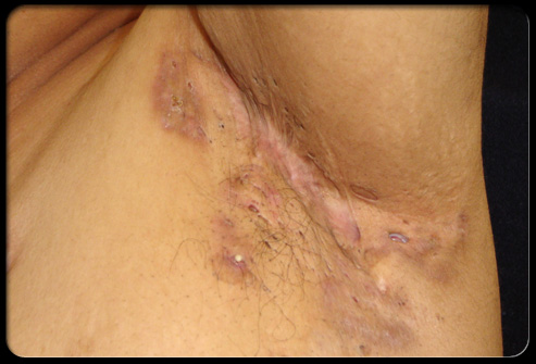
Microbiology & Pathogenesis
The infection is generally polymicrobial due mainly to aerobic and anaerobic bacteria of skin and proximal mucous membranes origin. Anaerobic bacteria alone or in combination with aerobic organisms were isolated from about two-thirds of patients. The predominant aerobic bacteria are S. aureus ( isolated from about a third of cases), GABHS, micro-aerophilic streptococci, and Pseudomonas aeruginosa. The most frequently isolated anaerobes are Peptostreptococcus, Prevotella, Fusobacterium and Bacteroides spp. 11
The anaerobes isolated from the patients are part of the flora of the oropharynx (Prevotella spp., Fusobacterium spp., Peptostreptococcus spp. and microaerophilic streptococci), gastrointestinal tract (Bacteroides spp., Peptostreptococcus spp.)11 and skin (Peptostreptococcus spp.) and presumably reached the HS lesions from these sites.
Diagnosis
The primary lesions are reddish-purple nodules that gradually become fluctuant and drain. Irregular sinus tracts with repeated crops of lesions are formed; reparative processes are only partially successful. The involved areas show a mixture of burrowing, draining tracts, and ciccatricial scarring. In some, HI is associated with acne conglobata or dissecting cellulitis of the scalp, that is often associated with spondyloarthropathy.
Management
Management is difficult and involves antimicrobial therapy, and moist heat locally to establish drainage in the initial phases of the infection. Large abscesses are surgically drained.
Gram's stain results may guide the clinician in selecting empiric antimicrobial therapy. However, the final choice of agents should be determined by the isolation of specific organisms, aerobes and anaerobes, and the results of sensitivity testing.
Initial empiric antimicrobial therapy should be effective against S. aureus as well as other potential aerobic and anaerobic pathogens. Antimicrobial agents active against S. aureus and anaerobic bacteria include clindamycin, a carbapenem, cefoxitin, and beta-lactamase inhibitor and penicillin combinations, and metronidazole with beta-lactamase-resistant penicillin. Cefoxitin and carbapenems also provide coverage against Enterobacteriaceae. However, agents active against Enterobacteriaceae (i.e., aminoglycosides, a quinolone, a fourth-generation cephalosporins) should be added when treating infections involving these bacteria.
PUSTULAR ACNE LESIONS
Acne vulgaris, a disorder of the pilosebaceous apparatus, is the most common skin disorder of the second and third decades of life.
Postular acne

Microbiology & Pathogenesis
Bacterial factors are important in the pathogenesis of acne. Acne is believed to be caused by Propionibacterium acnes.12 The improvement in acne patients treated with systemic antibiotics effective against P. acnes, as well as other organisms, support this concept.
The morphogenesis of acne lesions can be divided into two phases. The first phase is non-inflammatory, during which keratin accumulates in affected follicles producing whiteheads (closed comedones), which have very small orifices, and blackheads (open comedones) which have distended orifices. The second is an inflammatory phase during which a variety of inflamed lesions may develop from a proportion of comedones.
P. acnes is known to be associated with the inflammatory process in acne lesions,12 Propionibacterium spp. possess immunostimulatory mechanisms such as complement activation , stimulation of lysosomal enzyme release from human neutrophils, and production of serum-independent neutrophil chemotactic factors.
Organisms other than P. acne may contribute to the inflammatory process. These include Peptostreptococci and anaerobic Gram-negative bacilli such as Porphyromonas and Prevotella spp.13
Management
Antimicrobial therapy is a common adjuvant in the management of acne vulgaris. Topical or systemic antimicrobial agents effective against anaerobic bacteria including P. acne (i.e. clindamycin, macrolides and tetracylines) are benefital The empirical choice of antimicrobials may not always provide coverage for some of the resistant organisms that can be recovered from pustular acne lesions. Processing pustular specimens for aerobic and anaerobic bacteria can provide guidelines for adequate management of infected acne lesions.
REFERENCES
1. Finch, R.: Skin: and soft-tissue infections. Lancet. 1(8578):164–8, 1988.
1a. Brook I; The role of anaerobic bacteria in skin and soft tissue abscesses and infected cysts. Anaerobes 3:171-7, 2007.
1a. Brook I; The role of anaerobic bacteria in skin and soft tissue abscesses and infected cysts. Anaerobes 3:171-7, 2007.
4. Rosebury, T.: Microorganisms indigenous to man. New York
5. John, SD.: Trends in pediatric emergency imaging. Radiol Clin North Am. 37:995-1034 1999.
6. Jebson, PJ.: Infections of the fingertip. Paronychias and felons. Hand Clin.:547-55, 1998.
11. Brook, I. , Frazier, E.H.: Aerobic and anaerobic microbiology of axillary hidradenitis suppurativa. J Med Microbiol 48:103-5, 1999.
12. Burkhart CG, Burkhart CN, Lehmann PF. Acne: a review of immunologic and microbiologic factors. Postgrad Med J. 1999 ;75:328-31.
13. Brook I, Frazier E F, Cox M E, Yeager K J: The Aerobic and Anaerobic Microbiology of Postular acnes. Anaerobe 1:305 -7.1995.
Burn infections
.
Burn wound sepsis is a major cause of death among patients. Sepsis is characterized by progressive bacterial proliferation within the burned tissue, invasion into adjacent tissue, and systemic dissemination.
The surface of every burn wound is contaminated to some degree by bacteria.1 Therefore, most burn centers routinely monitor surface bacterial growth, allowing the determination of the effect of therapy and prediction of the bacterial strains that may be involved in sepsis.

Microbiology
The source of colonization of the burn wound usually is the patient's own endogenous flora as well as environmental organisms. These organisms can reach the wound directly through the skin or through the blood stream. Soon after a burn injury, surface cultures may reveal multiple organisms. Within three to five days, the wound will become colonized by one or two specific organisms that have survived the competition with other microorganisms or have proven particularly resistant to burn wound therapy.
A number of case reports describe the involvement of anaerobes in burn wounds. These reports were summarized by Murray
A report by Brook & Randolph 3 summarized data obtained from a prospective longitudinal evaluation study of the flora of burn surfaces in 180 children. total of 392 specimens were collected. Aerobic bacteria alone were present in 225 of 392 specimens (71%), and anaerobic bacteria alone were present in 26 (8%). Mixed aerobic and anaerobic bacteria were present in 68 burn specimens (21%). A total of 551 isolates (419 aerobes and 132 anaerobes) were recovered, accounting for 1.7 isolates per specimen (1.3 aerobes and 0.4 anaerobes). The predominant aerobic isolates were S. epidermidis, S. aureus, alpha-hemolytic streptococcus, Pseudomonas species and Enterococcus spp.. The predominant anaerobic isolates were: Propionibacterium acnes, anaerobic Gram-positive cocci and gram negative bacilli (including Prevotella and Porphyromonas sp. and Bacteroides fragilis).
Blood cultures were drawn from 45 children showed bacterial growth of one of each of the following isolates: S. aureus, Escherichia coli, Peptostreptococcus asaccharolyticus, and B. fragilis.
The number of isolates per specimen was higher in the oral and anal areas (3.2 and 2.8) than in the extremities and trunk (1.8 and 0.9). Gram-negative enteric rods and group D streptococci were recovered more frequently from the anal area. S. aureus, S. epidermidis, and P. acnes were more frequently recovered from extremities. Anaerobic gram negative bacilli and Fusobacterium nucleatum were more frequently recovered from the anal and oral areas. Specimens from burns of the anal and oral region tended to yield organisms found in the stool or mouth flora.4 Specimens obtained from burns in areas remote from the rectum or mouth grew primarily skin flora. With the exception of P. acnes, multiple anaerobic organisms usually were recovered from the anal and oral areas.
Wang et al5 studied 102 burn wound specimens obtained from 34 patients. Fifteen specimens from 8 patients showed growth of anaerobic bacteria. The predominant anaerobes were Prevotella melaninogenicas, Peptostreptococcus, Bacteroides fragilis, and other strains of Bacteroides and Peptostreptococcus. They were mostly discovered in electric burn wounds and burn wounds affecting the perianal and oral areas. Wounds with anaerobic infection usually appeared gaseous, necrotic and ischaemic with foul odour. Of 19 blood samples from 10 patients two were positive, one caused by Bacteroides and the other by mixed infection of Peptostreptococcus and Serratia.
Zhang6 analyzed 158 specimens from deep necrotic burn tissues and exudates under burned subeschar, and blood, and found that more anaerobes were isolated from deep necrotic tissues and foul smelling exudates of subeschar. Most isolates were mixed with aerobes (97%) and of the 43 strains of anaerobes, Clostridium accounted for 18, and Bacteroides for 17. Other isolates included Fusobacterium nucleatum. Peptostreptococcus sp., and Veillonella parvula.
Huang et a7 performed aerobic and anaerobic blood culture in 127 patients with extensive burns. The incidence rate of anaerobic septicemia was 20%, 61 anaerobes were isolated and 20 (77%) were mixed infection of aerobes and anaerobes. The predominant anaerobes were Peptostreptococcus (38%) and B. fragilis (36%).
Mousa8 who studied 127 patients recovered 377 isolates (239 aerobes, 116 anaerobes and 22 fungi). Anaerobic bacteria alone were present in four patients (3.2%). Mixed aerobic and/or anaerobic bacteria and/or fungi were present in 73 patients (57.5%). The predominant isolates were: P aeruginosa, S aureus, Bacteroides spp., Klebsiella spp. and Peptostreptococcus spp. There were 70 patients (55%) infected with anaerobic bacteria. The rate of recovery of anaerobes was higher in patients with open wound dressing (73%) than in patients with occlusive wound dressings (42%), (P < 0.01). Seventeen patients presented with septic shock, 15 of them (88%) yielding positive anaerobic cultures. Bacteroides spp. were isolated from 14 patients with septic shock, and were recovered from the four patients who had anaerobic infection alone.
Burn with a skin graft

Pathogenesis
The failure to use anaerobic methodology may account for the lack of recovery of anaerobes in many studies of the flora of burns. The recovery of these organisms from burns is not surprising, since anaerobes are part of the normal flora of the mucous membranes and skin.
The site of the burn can affect the isolated bacteria. anaerobes. . The burn wound can be susceptible to infection with anaerobic bacteria because of its necrotic, avascular qualities. Because anaerobes are predominant in the gastrointestinal and oral flora, they might colonize the burn wound, especially in burns adjacent to the oral and anal areas.
B. fragilis, a predominant anaerobe in the feces,4 was cultured most frequently from burns of the anal area. Prevotella melaninogenica, which occurs in stool as well as in the oral cavity, 4was most frequently recovered from the oral region. Most strains of B. fragilis and growing numbers of Prevotella, Porphyromonas and Fusobacterium sp are resistant to penicillin.
Burn

Management
Using appropriate aerobic and anaerobic microbiological techniques in monitoring the bacterial colonization of burns can help in selecting proper therapy if infection occurs. Local debridement of the wound should be done.9
In cases of invasive burn wound sepsis or septicemia, the wound should be examined, and a meticulous search made for subeschar abscesses. If no abscesses are found, multiple incisions through the eschar are made, to provide open drainage and to allow the antibacterial cream access to the deeper tissues.
General supportive measures should include evaluation of other sources of invasion (urinary tract infection, thrombophlebitis, pneumonia, etc.) as indications for intravenous fluid therapy and ventilatory assistance.
Broad-spectrum antibiotics should be administered parenterally until culture reports are available. This includes an aminoglycoside, a fourth generation cephalosporin effective against Pseudomonas such as ceftazidime for coverage of enteric Gram-negative rods, and a synthetic penicillin or cephalosporin for coverage of beta-hemolytic streptococci, enterococci, and S. aureus. If anaerobes are suspected, adequate coverage includes metronidazole, a carbapenem or the combination of a beta-lactamase inhibitor and a penicillin. Broad spectrum antimicrobial therapy should be used with caution, as it may have the untoward effect of predisposing to superinfection by yeast, fungi, or resistant organisms.
Burn patients have altered antibiotics pharmacokinetics due to the multiple burn related pathophysiologic changes.10 These differences must be considered in the selection of agent(s) and their optimal dosage. Patients who are not immunized against tetanus should have both active and passive immunization.
REFERENCES
1..Pruitt, B.A., Jr., Foley, F.D.: The use of biopsies in burn patient care. Surgery 98:292, 1969..
2. Murray
4. Gibbons, R.J. et al.: Studies of the predominant cultivable microbiota of dental plaque. Arch. Oral Biol. 9:365, 1964.
5. Wang, D.W., et al.: Anaerobic infections of burns.: Burns Incl Therm Inj 11:192-6, 1985.
6..Zhang, Y.P.: Anaerobic infection of burns. Chung Hua Wai Ko Tsa Chih 29:240-1,1991.
7.Huang, X., Ma, E., Gong, L.: Clinical analysis of anaerobic septicemia in 26 patients with extensive burn. Chung Hua Wai Ko Tsa Chih 33:752-3, 1995.
8..Mousa, H.A.: Aerobic, anaerobic and fungal burn wound infections. J Hosp Infect 37:317-23, 1997.
9. DeSanti L: Pathophysiology and current management of burn injury. Adv Skin Wound Care. 2005 ;18:323-32.
10..Boucher, B.A., Kuhl, D.A., Hickerson, W.L.: Pharmacokinetics of systematically administered antibiotics in patients with thermal injury. Clin Infect Dis 14:458-63, 1992.